Cell Envelope
The prokaryotic cells have a very complex cell envelope. It is composed of three layers, each with a distinct function.
- An outer Glycocalyx
- A tough Cell Wall
- An inner Plasma membrane
Glycocalyx
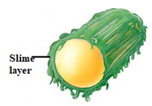
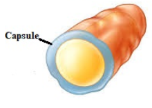
- Glycocalyx is a layer of polysaccharide (sometimes proteins) that protects the cell.
- The thickness and composition of glycocalyx differs in different prokaryotes.
- In some bacteria, it may occur as loose and mucilaginous sheath called the slimy layer.
- In some others, it may form a thick and tough outer covering known as the capsule.
- It is often associated with pathogenic bacteria because it serves as a barrier against phagocytosis by white blood cells.
Cell wall
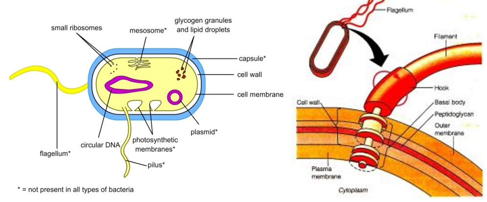
- The cell wall is the structure that determines the structure of the cell and prevents the cell from bursting or collapsing.
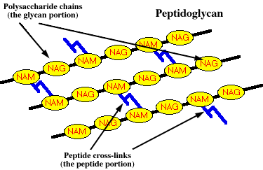
- It is made of a complex substance called Peptidoglycan (or murein).
- Peptidoglycan is a polymer consisting of interlocking chains of two types of subunits, namely
- NAG – (N – acetyl glucosamine) and
- NAM – (N – acetylmuramic acid).
- Glycosidic bonds link these monomers to form chains of peptidoglycan subunits.
- These individual chains are then joined to one another by means of peptide.
- It provides tremendous strength to the cell wall.
- In Archaebacteria, the cell wall is composed of pseudomurein
- In mycoplasma, there is no cell wall.
Plasma membrane:
- The plasma membrane is a selectively permeable membrane.
- It is a bi-lipid layer which helps in the interaction of cell with the external environment.
- There are numerous proteins moving within or upon this layer that are primarily responsible for transport of ions, nutrients and waste across the membrane.
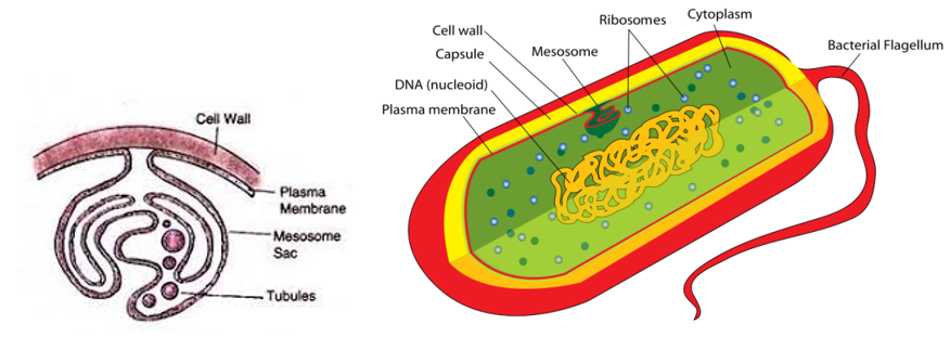
- Mesosomes
- The plasma membrane appears to be infolded at more than one point.
- Such complex infoldings are called mesosomes.
- These extensions are in the form of vesicles, tubules and lamellae.
- These are believed to be the principal location of respiratory enzymes. Thus, they help in respiration, increases the surface area of the plasma membrane and enzymatic content.
- They are also involved in cell wall formation, DNA replication and distribution to daughter cells.
- In some prokaryotes like cyanobacteria, there are other membranous extensions into the cytoplasm called chromatophores which contain pigments.
Gram positive and Gram negative bacteria.
Based on the differences in cell membrane, and the manner in which they respond to the staining procedure developed by Christian Gram, bacteria are classified as Gram positive and Gram negative bacteria.
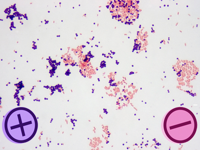
- Gram positive bacteria stain violet due to the presence of a thick layer of peptidoglycan in their cell walls.
- Peptidoglycan absorbs the stain ‘crystal violet’.
- Alternatively, Gram negative bacteria stain red.
- This is due to the thinner peptidoglycan wall, which does not retain the crystal violet during the decoloring process.
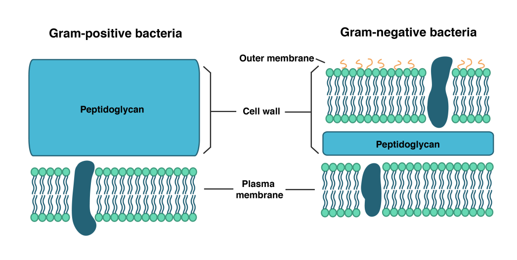
- The bi-lipid layer in gram negative bacteria is the source of lipopolysaccharide (LPS) in these bacteria.
- LPS is toxic, and is called endotoxin.
- LPS is absent in Gram positive bacteria
Cell Wall Appendages
Pili
- These are hollow tubular structures made of protein subunits known as Pilin.
- A specialized pilus, called sex pilus, allows the transfer of genetic material from one cell to another.
Fimbriae
- The fimbriae are small bristle like or hair like fibres sprouting out of the cell.
- In some bacteria, they are known to help attach the bacteria to rocks in streams and also to the host tissues.
Flagella:
- Flagellum is the principle locomotors organ in prokaryotes.
- These are long appendages consisting of three components, namely a basal body, a hook and a shaft.
- These are made up of protein subunits called flagellin.
- Prokaryotes may have one, a few or many flagella in different positions on the cell.
